Support Us
Donations will be tax deductible
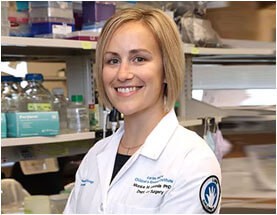
Monica M. Laronda, PhD, Assistant Professor of Pediatrics (Endocrinology), provides an overview of her research work in endocrinology, reproductive biology, and 3D printed ovaries.
Laronda is passionate about research and she has a keen interest in the reproductive biology and endocrinology that forms the foundation for the the development of treatments to protect or restore hormone function and fertility. Laronda received her PhD from Northwestern University and her Postdoctoral Fellow work was done at Northwestern University’s Feinberg School of Medicine (Obstetrics Gynecology).
Laronda talks about their main patient focus: childhood cancer survivors whose treatment or disease may have rendered them infertile later in life. She talks about bioprosthetic ovaries and the uses of 3D printing. As she explains, their early work has shown success in mice, and she discusses what that means for future research. The PhD explains how egg cells work, and the natural decline that happens over time, as well as how disease can affect them. She discusses the complexity of the ovary, and her lab’s research in which they investigated biochemical differences, etc. to assist in the development of the best bioprosthetics. She discusses transplanted tissue and how long it can last once transplanted.
The endocrinology expert explains their scaffolding models and her team’s hopes for further study. And Laronda provides information on the micro-environment, scaffolding proteins, etc. that play a role in cellular functions.
In this podcast you’ll learn:
How 3D printing is helping to advance bioprosthetic development
The importance of primordial follicles
How disease can impact fertility
Richard Jacobs: Hello, this is Richard Jacobs with the future tech and future tech health podcast. I have Monica M. Laronda. She’s an assistant professor of pediatrics and endocrinology at Northwestern. I’m talking about a bioprosthetic ovary that was created using a 3D printing method, which you’ll get into, so Monica, thanks for coming.
Monica M. Laronda: Yeah, thanks for having me. I’m always excited to talk about our research.
Richard Jacobs: Maybe it’s obvious, but what’s the reason to try to recreate a 3D printed ovary?
Monica M. Laronda: Our main patient population is childhood cancer survivors. So, there are children that either has a disease or a treatment for their disease that would render them infertile later in life and so they would have a reduced reproductive capacity and reduced sex hormone function. And so developing a bioprosthetic ovary or a 3D printed ovary is one way that we’re trying to advance their regeneration options.
Richard Jacobs: Hmm. So I mean, how far along in this process are you, have you been able to construct an ovary and have any functionality at all or is it still really early on in the process?
Monica M. Laronda: Yeah, we’ve been successful in mice. So we’ve been able to 3D print a scaffold that supports the ovarian cells that are important for function. So the potential egg cells or the oocytes and the surrounding supportive hormone-producing cells together that’s called an ovarian follicle. And it’s critical. One of the things that we discovered was that it was very critical to have this spheroid structure, this cell aggregate structure maintained in a particular 3D printed geometry that would allow them to grow and mature and ovulate. First, we found that it ovulated in culture. And then we were very excited to then transplant these bioprosthetic ovaries and mice whose ovaries were removed mate them and they were able to produce pups. So their hormone and fertility were restored with this transplant.
Richard Jacobs: Oh, wow. I haven’t really thought about ovaries very much. I don’t think most people have. But in a normal ovary, like I would guess you’d have the cells dividing normally. And then how is the actual egg cell produced? Is it a differentiation of a different cell type that exists in the ovary and that becomes an egg cell or how does an egg cell come into being?
Monica M. Laronda: Yeah, that’s a really good point and it brings me to a very important factor about female fertility, which is that our oocytes are a finite resource. They are actually, once we’re born, we have the most number of potential egg cells that we’ll ever have. And it just declines from there until natural menopause. Or until something destructive comes along either a genetic mutation or a disease or a treatment that would affect those cell numbers. The oocytes are actually paused in meiosis, which if you remember from biology classes, not the same as mitosis or they’re not doubling. They’re not, they’re not copying themselves. They’re trying to create like half their genetic material so that the two halves can be combined and develop into an embryo in a new human being, the egg, and the sperm. So they’re frozen that way at birth.
Richard Jacobs: Okay. So from what you’ve observed, it is true that by the time an organism is born, it has all the egg cells it’s going to have and then they hang out, they’re not doing much. And then according to some hidden timing schedule, one or more of them every month or every gestation cycle will now suddenly undergo meiosis and create what?
Monica M. Laronda: Yeah, so I think the egg expert colleagues that I have would want me to point out that the egg is not just hanging out there doing nothing. That is actually an important contributor to the process. But in some sense, it’s paused in meiosis, but it does really grow and expand. And that’s one of the aspects that we have to consider when we create the 3D printed scaffolding that the oocyte itself and the cells surround it will expand to over 600 times their original size. And so it just, you just have to have this dynamic structure that is able to hold them. And so as it’s growing it kind of prepares itself and it’s kind of a black box as to actually what happens in some respects which makes doing this sort of research really fascinating.
But it readies itself to complete meiosis upon ovulation. And then the sperm will combine its half of the DNA to then develop, first create a zygote and then develop into an embryo. If those implants, then it becomes a fetus and a whole unique being.
Richard Jacobs: Have you been even considering, and I’m realizing how little I know. So when you 3D print an ovary and you said you’ve done it in mice. I guess you are 3D printing the egg cells as well, right? So, when a person is born are their egg cells immature in the age with the person, and then when the person reaches a certain age, then the cells are ready according to some clock to start being candidates for meiosis and traveling down the fallopian tubes, I mean, or the egg cells like ready at all times to be harvested.
Monica M. Laronda: Yeah, that’s a great question. And I think what might help is if I create that link between the patient and how we’re going to use like the strict generative medicine type of technology. So when a child is diagnosed with cancer and they’re going to receive this treatment that puts them at over 80% risk of developing infertility, they qualify for our ovarian tissue crier preservation procedures that we have at Lurie children’s hospital. I co-direct the fertility and hormone preservation and restoration program with my colleague, Aaron Raul, who’s a pediatric surgeon and her and I offer this clinical procedure to patients. And so an ovary is removed, one ovary is removed ideally prior to their initial chemotherapy or treatment. So before it would be exposed to these potentially harmful effects.
It’s processed and then cryopreserved. So there’s been several patients who have had offspring. So they have gotten pregnant and had live births from ovarian tissue that was transplanted after it had been cryo-preserved in this same way that we do here in the clinic. However, there are many cancer patients that have metastatic disease. And so they wouldn’t qualify for this transplantation procedure because we don’t want to like give them back the disease that they just survived. And so what we do with the tissue is we isolate out the cells that we want. So we want the oocytes from that patient because it will have their biological material, which is very important for them to pass on to their offspring. And we’ll also harvest the hormone cells. And we’ll put that into a 3D printed scaffolds that we make in the lab. And so then the transplant patient happens like it normally would have. But this time we know that there is no metastatic cells in there. And hopefully, we can even make a better version of what exists with the tissue today. So the scaffolding itself is what’s actually printed and it’s at a specific architecture to hold those ovarian follicles, which is the potential egg cells in the hormone-producing cells. And we seed those into the scaffolding.
Richard Jacobs: Oh yeah, I got that. But I guess my question was if I have someone that’s five years old and I cryopreserve one of their ovaries and now they are 25. Everything looks good. That puts ovary back into them. They can have kids within what time period?
Monica M. Laronda: Yeah, that’s a good question. So, most of the live births that have happened are from adults or post-pubertal children that have been cryo-preserved when they were postpubertal and then transplanted back when they were adults and wanted to get pregnant. So there’s only been only two cases where this tissue is transplanted back that was preserved prior to them going through their full pubertal transition. So we hope that it works the same. We know from animal studies and from some human studies that we can get and eggs produced from these immature primordial follicles that are collected from pediatric patients. But we’re actually doing a large research study to figure out the quality and the rate at which this can happen. Because you’re right, it’s coming out of a person who doesn’t have that hormonal cycle, which is what activates those follicles and matures those oocytes into good quality eggs. That happens through puberty and after puberty in adult women.
Richard Jacobs: Yeah. I wonder if anyone’s done a longitudinal study on oocytes and looked at them over a person’s lifetime. Cause you know, because like, for instance, a woman’s cycle has timing, definite timing to it. So you wonder if the woman’s first cycle matures all her eggs to a point where they’re candidates to the travel and the flow takes X number of cycles.
Monica M. Laronda: No, no, no. There are a few oversights that are recruited at a time and then one very special one is ovulated and that happens between 28 and 32 days in a normal cycling female. But not all of them are all recruited.
Richard Jacobs: Oh, so once you’re chosen, you’re picked out of the bunch. That’s it. It’s either go or no go.
Monica M. Laronda: Yeah, it’s either they die off or they’re ovulated, but it’s a cluster of them at a time. But it depletes like we’re born with about 300,000 and then it depletes down to about a thousand per ovary once we reach menopause. And so there are several recruited that don’t make it to eggs. But not all of them are recruited at once. It happens over the decades.
Richard Jacobs: Well, okay. Okay. I gotcha. But again, a study on the ones that are just hanging out in the freezer, essentially are hanging out in the ovary, not matured yet or not picked. And what happens to them over time?
Monica M. Laronda: Well, there’s been some studies definitely because we do the way that we do assist reproductive technology with like IVF, in vitro fertilization, many families have to go through that process where the woman has an increased hormone regimen to recruit more ovarian follicles to grow. And then we’re capturing them before they get ovulated out of the ovary. And then using those in the lab to make embryos once combined with sperm or to freeze them for later. And so we do use some of those eggs that wouldn’t normally be ovulated to create embryos and to create beings with the IVF’s and things like that. But we are studying, I think where there’s a lack of research is the pre-pubertal and through the pubertal transition, which I think because I’m in the department of pediatrics, I’m very interested in like what happens during that transition and how does the ovary change and how does the capacity of the follicle to be good quality eggs change over time.
Richard Jacobs: Can you tell which eggs are going to be next on deck to be developed? Like the way the eggs are stored, are they stored in like a regular like honeycomb pattern? And let’s say the ones in the quote-unquote top or nearest the exit are the ones that are most likely to be taken for the next batch?
Monica M. Laronda: There’s a lot of theories out there. So the ones that are kind of the bank, we call them the bank of eggs or potential eggs those primordial follicles they’re in a more rigid part of the ovary. So they’re in what’s called the cortical region. And then they migrate into the middle of the ovary before they expand and then reach the edge again where they then ovulate out. But the growth happens within the middle, which is really interesting. And the bank stays on the outer perimeter. So there’s a lot of theories as to which ones are going to be recruited next based on which ones were formed first. In the embryo which ones underwent meiosis first or completed? The first part of meiosis to be quiescence. And then also where they were located in the tissue, not just in the cortex, but where they next closer to the kidney or further away, for example. Yeah, there’s a lot of theories out there, but I don’t know that answer.
Richard Jacobs: I’m sorry I’m asking this stuff that’s coming to mind, but I just realized, Oh, this is something that’s very complex and I haven’t really thought about it at all, but most people haven’t. So that’s why I’m asking these questions because all that stuff’s popping into my head now and I’m like, wait a minute.
Monica M. Laronda: Yeah. And this is why using 3D printing is interesting and I think useful in this particular case because the ovary is complex and compartmentalized and we’re publishing as a study soon from my lab where we investigated all of the structural proteins of the ovary and mapped it spatially throughout the ovary to define these not just rigidity differences, but biochemical differences that might actually affect how follicles behave. And in attempts to make the best possible ovarian bioprosthetic.
Richard Jacobs: So this is not necessarily a permanent solution but is this maybe a short term one where you said someone’s had childhood cancer, you were able to preserve at least the egg cells, if not the whole ovary itself and now is your best hope to give them a window, like a three to a five-year window or something where they could have natural-born children, but that’s about all you’re going to get. Or like what’s the goal of the project?
Monica M. Laronda: Yeah, that’s a great question. So when we transplant back ovarian tissue the average age that tissue lasts, and so it’s quite often about 20% of the tissue is put back at a time, it lasts about two to five years, but sometimes it lasts longer, sometimes it lasts like 12 years. And this is based on the type of ovarian sex hormones that are produced and testing that within the blood. And so our goal is to have long-term transplants and we are able to do better than the natural ovarian tissue that exists in those types of transplants just because we might be able to control the environment a little more with the scaffolding with the 3D printing. And so we’re hoping to get a long-term transplant. But I think short term restoration of fertility is definitely one of our key like targets when we first start moving these into the clinic. First looking for a hormonal restoration and then seeing if we can get some short term, year or two ovulation to help them have a biological child.
Richard Jacobs: I figured. Yeah. So what are you trying to 3D print, the whole ovarian structure or just the last stage of it? The last bank parts where they’re ready to be selected for maturation?
Monica M. Laronda: Yeah, that’s a good question. So that what our first focus is to do the cortical region. So exactly right. The region that the OSI is banked. And this is because generally we take out one ovary to cryopreserve it and the other ovary remains. And so we wait until that ovary essentially goes through early menopause. We call it ovarian insufficiencies. And then we transplant it back onto that remaining ovary. And so the rest of that tissue is there. Whether or not it’s an ideal organ to put it on is another thing that we need to test because we’re sure that a post-menopausal ovary isn’t the perfect environment, but the first attempts that we’re going to do are to transplant it back on, onto that remaining ovary. And so we’ll just need to make that bank a cortical region to how’s the primordial follicles?
Richard Jacobs: Well, the reason I ask you just so complicated, you know, good luck if you can even do a small part of it.
Monica M. Laronda: Yeah. You’re really getting at like basically our stepwise strategy of our first lines of successes. And then, of course, there are the ultimate goals of recreating the entire organ and it functions for a longer time than the natural ovary. That would be ideal. For women to not have to go through menopause and then stay that way for about half their life at this point. So that would be awesome if we could recreate the whole ovary.
Richard Jacobs: Well, what happens to the ovaries and menopause? I mean, do they stop growing? I mean, do they still continue apparently as normal? It’s just that the oocytes aren’t formed at the end or does the whole thing shut down? Like what’s been observed?
Monica M. Laronda: Yeah, Basically the whole thing shuts down. So there are gonadotropins that control follicular genesis and respond to hormones that come from the ovary. So those are off-kilter and response and the ovary just stops producing estradiol. I mean, it’s a decline, so it’s not an immediate-type thing. Though it might feel that way for a lot of women where they all of a sudden just have all of these symptoms of complete estrogen withdrawal and no more progesterone. And then there are, of course, other things that the ovary produces. But there is, interestingly, there is still a bank left. As I said, there are about a thousand primordial follicles left. It’s just not enough for the hypothalamic-pituitary-gonadal axis of hormones to continue on with that cycle that would then trigger ovulation and then result in more estradiol produced and it’s a whole cyclical effect.
Richard Jacobs: This may be an over-simplification, but I see it as three stages. There is like the bank stage to the primordial part where they hang out. And you said that gets down to be about a thousand and then it just kind of sits there, I guess forever. Then there’s the growth, the growth part in the middle where they grow huge 600 times the size. Then there’s the last stage now where they’re prepped and shoved out the door to, you know, preconception. So in a postmenopausal woman, the first stage is still there. There’s still the bank itself sitting there, but they just not progressed to the second stage I call it, you know, where they start to grow and expand and develop or is it just the third stage that’s effective.
Monica M. Laronda: No, it’s the second stage so they’re not recruited to grow. And the growing ones actually produced the Ash dial. And then the post-ovulatory follicle creates this small gland that produces progesterone and then regresses. So the hormone production happens at what you’re identifying as the second and third stages.
Richard Jacobs: Okay. That’s good.
Monica M. Laronda: Yeah, so the bank of follicles exists from birth to death but are just kind of hanging out until they’re recruited and then they stopped being recruited.
Richard Jacobs: Gotcha. Okay. Yeah, I just wanted to understand it. I appreciate it. So you’re making a scaffolding, so I guess you’re modeling that on what the last third or the last part of the ovary looks like, that houses the cells that are just about ready to, again, I just call it the head out the door and become candidates for fertilization. That’s what you’re trying to recreate with your scaffolding is just that section.
Monica M. Laronda: We’re actually trying to create the first part, the bank, I’m hoping that they’ll grow, they’ll be activated within the remaining ovary. And that the scaffold will be dynamic enough. Like it was in the mouse model actually to withstand those changes in size and actually just be remodeled by the follicles and the other cells that they recruit within the scaffold itself to actually reform and kind of remake that tissue. So we’re kind of assuming that this is what happens in development and this is what I’m excited to discover. And some of our future research studies but we think that the pre-pubertal ovaries is more like the whole cortex. So once those follicles are triggered to grow, then a Medulla type forms where it gives it space and we’re hoping to kind of recreate that with the 3D printed scaffold and with the help of the remaining ovarian tissue. I hope that makes sense.
Richard Jacobs: So you’re saying that science doesn’t know the morphology of the ovary and how it changes from birth through puberty to the adult stage. We know what it looks like in adults.
Monica M. Laronda: We know what it looks like in the prepubertal ovary. We see them all the time, but we don’t know how the environment, the microenvironment, the scaffolding proteins, and the stromal cells that are around it, how that changes. And that’s what we’re studying in the lab. But in the mouse model, we were able to create this 3D printed scaffolds where it houses the small follicles, but then once they were triggered to grow because it’s made from gelatin, which the cells can interact with and digest and break away it, they just reformed an ovary. They recruited vessels when it was transplanted and it ovulated right through it. So we’re hoping that a similar thing will happen when we create it for large animal models. And then for humans,
Richard Jacobs: I guess we don’t understand the cell to cell signaling within the ovarian structures with the surrounding microenvironment then with the surroundings, other organs and the rest of the organisms. So there’s probably multiple levels of signaling going on back and forth. And it sounds like we’re really don’t understand almost any of that yet.
Monica M. Laronda: Yeah. And it’s really a fascinating part of biology. I think that if you just give the cells the right environment, that they’ll behave how they need to and that there might be some form of control that we can create within the 3D printed scaffolds which is exciting that we might be able to design something that’s even better.
Richard Jacobs: So have you tried different orientations and morphologies of the scaffolding given that the body’s going to do what it’s going to do anyway and remodel the thing, have you found more advantaged structures or morphologies to start out with that seemed to work better for them?
Monica M. Laronda: Yeah, absolutely. We did. So we, at first, because they’re round, I thought that we could print these round pores and just kind of slot them in and that would be good. But first, it was very hard to actually get the follicles into something that was meant to be about the same size and they weren’t captured in the pores like they weren’t held within different layers of the scaffolding cause you imagine we want to fill it to capacity and there are several layers to this 3D printed structure. It just really didn’t work. And so we tried angles then. So we tried 90-degree angles and 60-degree angles and 30-degree angles. And it turned out that 60-degree angles and 30-degree angles were the best ones to support the ovarian follicles. And the 60-degree angle was the one that we actually use for the 3D printed scaffolds because not only was it supportive of the follicle, but it also gave us enough space to put more of them in. One piece of a scaffold. So it was quite interesting to kind of go through that and to figure out that we needed to do the angular and advancing offset 3D printing design in order to hold something that was round.
Richard Jacobs: If you’re going to 3D print this and make it, why not study how the ovary is formed in utero when the embryo is developing. I mean, if you could picture it somehow, if you could see it and see how it’s developed, maybe it’s developed like radially outwards from a central point. Maybe it’s built up, I don’t know bilaterally on two sides. It’s probably not built up at all like a 3D printed way and layer by layer. Maybe if you’re able to figure that out and emulate that, you’d have more success in making it,
Monica M. Laronda: Yeah, I see your line of thinking. The problem is on this, the cells of the ovary and as its developing can be singular so they can be isolated cells and they can migrate. So there kind of long and flat and what you would traditionally think of like a fiber plastic-type cell. But the OSI in, once we’re born, it can’t be uncoupled from the granulose cells. So we need to maintain that sphere. And so it has to be, the scaffolding itself has to be something that has a pocket to hold this sphere together. Does that make sense?
Richard Jacobs: Okay. Yeah, I gotcha. Well, you’ve chosen yourself a tough challenge, but it’s a good one. So where are you at specifically with this? So you said it’s worked in mice. Has the clinical trial in the works for this in people, or is it not at that stage yet where it’s working well enough to do that?
Monica M. Laronda: Yeah, it’s not that stage yet where we’re testing animal models first. And we’re doing some investigations, like I mentioned, of what the microenvironment looks like in both larger animal models and some ovarian tissue that participants donate, which we’re extremely grateful for. But we’re looking more into the microenvironment because we think humans aren’t mice. And so we expect that it will be more complicated and then using a gelatin egg for the 3D printed scaffolds, but we’ll need other types of proteins and biochemical cues for the follicles for larger animal models.
Richard Jacobs: Well I think just the uniqueness of your gelatin model that allows the cells to migrate and be the host to orchestrate them as it needs to. I mean, I think that’s a big innovation and I’m sure other people are going to try to 3D print new scaffolds would benefit from that kind of scout scaffolding themselves.
Monica M. Laronda: Yeah, I think so. Especially anything that needs to be maintained in an aggregate structure. The way that our follicles do. I think that this type of design was an important piece to work out and why not use our favorite animal model, mice. Plus we were able to use them genetic manipulations to show that pups that were born were from the transplant and not from the mom that gave birth to them if that makes sense. We used a green fluorescent protein, which is always fun.
So the pups came out fluorescing green. But yeah, I think that was an important part of the design. And so to work out the architectural part.
Richard Jacobs: Yeah, definitely. So what animals are next? You know, what animals can you work with?
Monica M. Laronda: Yeah, we’re thinking of, so we have a few animal models that we’re working on for different aspects of this. So we were able to receive the poor sign and bovine, so picking cow tissue from slaughterhouses which is great for us to use because their ovary compartments are more like humans than mice are. So that’s what we’re using to study, kind of the architecture and the design. And then we of course always go back and confirm it with human tissue. But as far as transplants we’ll have to see what type of animals will be the best option for those.
Richard Jacobs: Okay. Well, no, this is really fascinating. So what’s the best way for people to learn more? I don’t know if they could have read scientific papers, but how can they look and see what you’re doing and learn more even if they’re not like an egg spirit.
Monica M. Laronda: Yeah, yeah. So I definitely have a faculty webpage at both Northwestern University and Lurie children’s, our clinical website is luriechildrens.org/fertility and my personal lab website where you can read little bios of the lab members that work in my lab and here are some updates of what we’re doing is larondalab.org.
Richard Jacobs: Okay. Very good. Well, I appreciate you coming in. It’s been a really cool conversation and I hope I haven’t driven you crazy with the questions.
Monica M. Laronda: Oh, no, no, no. It’s always fun to talk about our research. Especially when I feel like I’m informing people about things that they don’t normally think about. So thank you.
Podcast: Play in new window | Download | Embed

Under the new presidential administration, public health policy is taking a new direction. With RFK Jr. now serving as the U.S. Secretary of Health and Human Services, significant… Read More

In today’s episode, we dive into the realm of neurological wellness with Adam Schell, the owner and founder of Brain Supreme. Brain Supreme is a transformative microdose supplement… Read More

In today’s episode, we connect with Dr. Alan Breen to discuss motion analysis and musculoskeletal modeling and how they relate to the treatment of spinal disorders. Dr. Breen… Read More

In today’s episode, Dr. Karen DeCocker, PMHNP, DNP, CNM, joins the podcast to discuss the use of ketamine to treat depression and various other mental health issues. Dr.… Read More

Can bioengineering improve the health, vitality, and longevity of human beings? What would the future look like if the medical system were about preserving health rather than just… Read More


Subscribe to Our Newsletter
Get The Latest Finding Genius Podcast News Delivered To Your Inbox